
On this month's Morbidly Fascinating Page:
Utrecht Hospital and some of its patients from the late 19th Century
IN THE ARCHIVES:
Dinosaurss to Birds
Ghosts of Alcatraz
Hansen’s Disease
Accidental Photos
Deaths on Mt. Everest
Titanic Graveyard
Time Travel
A set of haunting medical portraits reveal the extreme symptoms suffered by patients at one of Holland's oldest hospitals in the late 19th century.
The photographs, which date back to the 1890s, were part of medical records collected for patients at the Utrecht University Hospital in the Netherlands. The hospital, which is now known as University Medical Center Utrecht, is one of the country's oldest clinics and was founded alongside Utrecht University in 1636. These portraits provide an insight into the early days of medical photography.
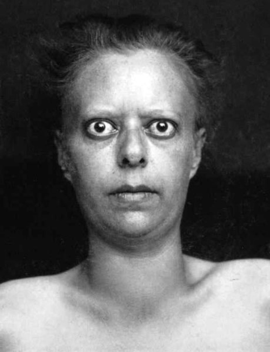
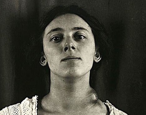
This patient was seen for hyperthyroidism, where the thyroid gland produces too much of the thyroid hormones. Hyperthyroidism is a condition that occurs when the thyroid gland is producing excessive thyroid hormones. The thyroid gland is a small butterfly shaped gland located in front of your neck. It has two lobes, the left and right and is connected by the isthmus. Bulging or protruding of one or both eyes is called proptosis or exophthalmos. Exophthalmos is caused by Graves disease, a disorder causing overactivity of the hyperthyroidism.
Today's treatments: Radioactive iodine. Taken by mouth, radioactive iodine is absorbed by your thyroid gland, where it causes the gland to shrink. Anti-thyroid medications. Beta blockers. Surgery (thyroidectomy).

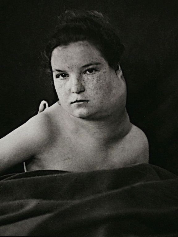
A young girl suffering from a Melanocytic nevus. These dark-colored, often hairy, non-cancerous patches of skin usually continue to grow as the child grows. The skin of the nevus is often dry and prone to irritation and itching (dermatitis). Excessive hair growth (hypertrichosis) can occur within the nevus. There is often less fat tissue under the skin of the nevus; the skin may appear thinner there than over other areas of the body.
Today's treaments: Melanocytic nevi can be surgically removed for cosmetic considerations or because of concern regarding the biological potential of a lesion. Melanocytic nevi removed for cosmesis are often removed by tangential or shave excision. Punch excision can be used for relatively small lesions.
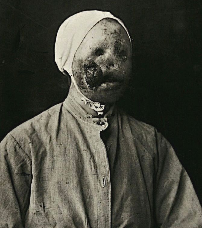
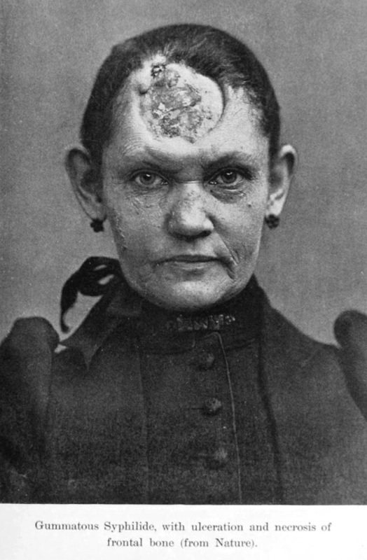
Two photos of women suffing from Gummatous syphilis, which is characterized by granulomatous lesions, called gummas, which are a center of necrotic tissue with a rubbery texture. Gummas principally form in the liver, bones, and testes but may affect any organ, including the skin. These are severe cases of Stage 4 syphilis.
I am not entirely convinced that the first woman suffered only from syphillis, even though the photo was marked as such. I wonder if she was also affected by something like leprosy. The second photograph was not in the Utrecht collection but is used here to demonstrate a single lesion to show how much damage syphillis alone could indeed cause.
Today's treatment: When diagnosed and treated in its early stages, syphilis is easy to cure. The preferred treatment at all stages is penicillin.
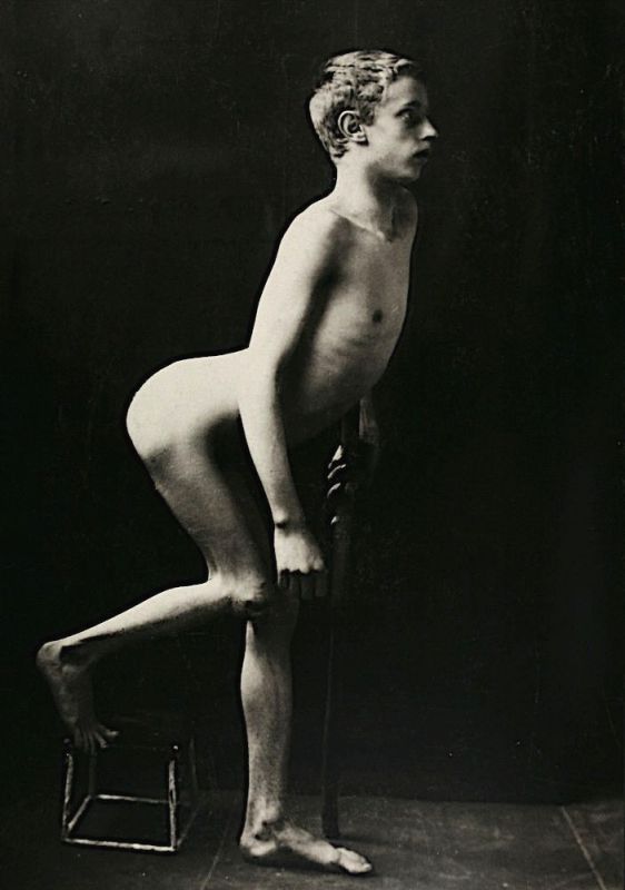
This man was suffering from Lordosis, a type of deformity which can create a curvature of the spine. This person's curve was probably caused by one of these things: Spondylolisthesis, a condition in which a vertebrae, usually in the lower back, slips forward; or Discitis, or inflammation of the disc space between the bones of the spine most often caused by infection.
Today's treatment: Medication to reduce pain and swelling. Nutritional supplements such as vitamin D. Daily physical therapy to strengthen muscles and range of motion. Braces in children and teens. Surgery is used in severe cases.
A patient suffering Hibernoma, a rare benign tumor of the neck. It is is an uncommon benign fatty tumor that arises from the vestiges of fetal brown fat. It is named “hibernoma” because of their resemblance to the brown fat in hibernating animals
Today's treatment: Surgical removal.
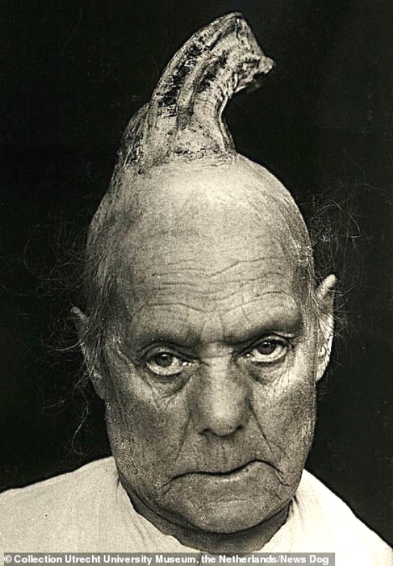
The cutaneous horn appears as a funnel-shaped growth that extends from a red base on the skin. It is composed of compacted keratin (the same protein in nails). Squamous cell carcinoma is often found at the base. A cutaneous horn is more common in older patients, with the peak incidence in those between 60 and 70.
Exposure to radiation from the sunlight may be one of the causes. Another possible cause is having viral warts caused by human papillomavirus. It's estimated that about half of cutaneous horns appear on top of, or because of, skin cancer or precancerous skin lesions.
Today's treatment: Options include topical chemotherapy, cryosurgery, electrodesiccation and curettage, surgical excision, Mohs micrographic surgery, radiotherapy, and photodynamic therapy.
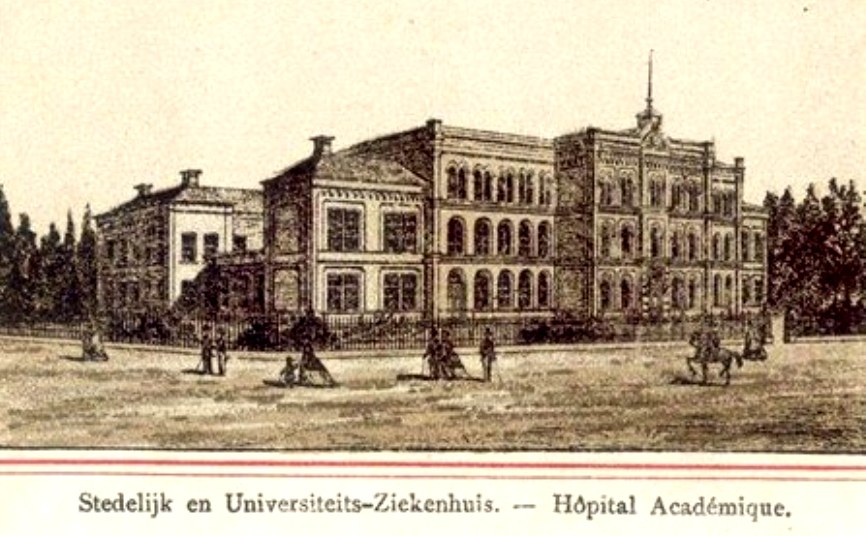
Drawing of 1890 Bartholomeus Gasthuis hospital (Utrecht) in the late 19th Century (above).
The Bartholomeus Gasthuis is one of the oldest properties in the city, and was the very first hospital in the Netherlands. Sometimes it is referred to as Utrecht Hospital since that is the name of the town where it is located.
History
The history of the Bartholomeus Gasthuis dates back to the 14th century. The nursing home was founded in 1367 and dedicated to St Apollonia. In 1378, it formally became a residence for poor people with chronic illnesses and has, over the past 600 years, served unbrokenly as a care home for elderly Utrecht residents.
Through the centuries, the hospital has undergone multiple renovations in order to comply with the requirements of new laws and regulations. For example, much changed after the Second World War with the emergence of an effective system of social care. The charitable nature of the hospital gave way to modern views on care for the elderly, with key principles such as increased privacy, more amenities and better care, with the result that the hospital came to feel more and more like a real home.
The photographs
The photographs on display came from the archives of Utrecht`s university hospital – records of medical disorders, photographed since circa 1890.
In the early days of medical photography clinical standards had yet to be formulated for photographic images. Consequently, many photographs are more poignant and beautiful than they are scientific. Light, space and patients` complete submission to doctors and photographers evoke feelings of compassion, surprise, embarrassment and amusement rather than disgust or scientific curiosity.
The exhibition Utrechtse Krop in De Kabinetten van De Vleeshal centred around the appeal of illness and the fragility of our physical being.